F- CLINICAL ASPECTS OF SPINAL CORD INJURIES
493
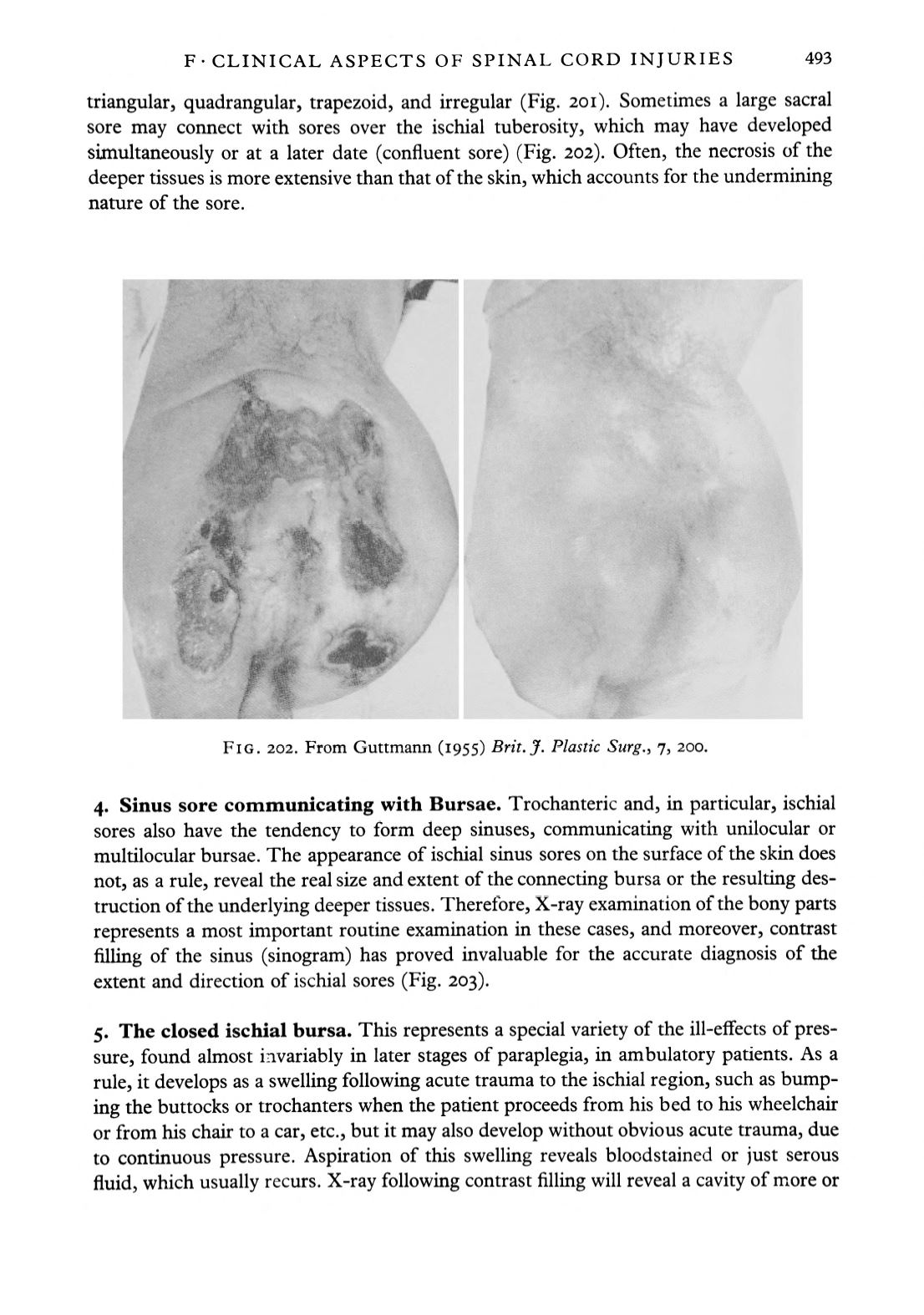
triangular, quadrangular, trapezoid, and irregular (Fig. 201). Sometimes a large sacral
sore may connect with sores over the ischial tuberosity, which may have developed
simultaneously or at a later date (confluent sore) (Fig. 202). Often, the necrosis of the
deeper tissues is more extensive than that of the skin, which accounts for the undermining
nature of the sore.
FIG. 202. From Guttmann (1955)
Brit.J. Plastic Surg.,
7, 200.
4. Sinus sore communicating with Bursae.
Trochanteric and, in particular, ischial
sores also have the tendency to form deep sinuses, communicating with unilocular or
multilocular bursae. The appearance of ischial sinus sores on the surface of the skin does
not, as a rule, reveal the real size and extent of the connecting bursa or the resulting des
truction of the underlying deeper tissues. Therefore, X-ray examination of the bony parts
represents a most important routine examination in these cases, and moreover, contrast
filling of the sinus (sinogram) has proved invaluable for the accurate diagnosis of the
extent and direction of ischial sores (Fig. 203).
5. The closed ischial bursa.
This represents a special variety of the ill-effects of pres
sure, found almost invariably in later stages of paraplegia, in ambulatory patients. As a
rule, it develops as a swelling following acute trauma to the ischial region, such as bump
ing the buttocks or trochanters when the patient proceeds from his bed to his wheelchair
or from his chair to a car, etc., but it may also develop without obvious acute trauma, due
to continuous pressure. Aspiration of this swelling reveals bloodstained or just serous
fluid, which usually recurs. X-ray following contrast filling will reveal a cavity of more or


