C- DISLOCATIONS OF THE VERTEBRAL COLUMN
151
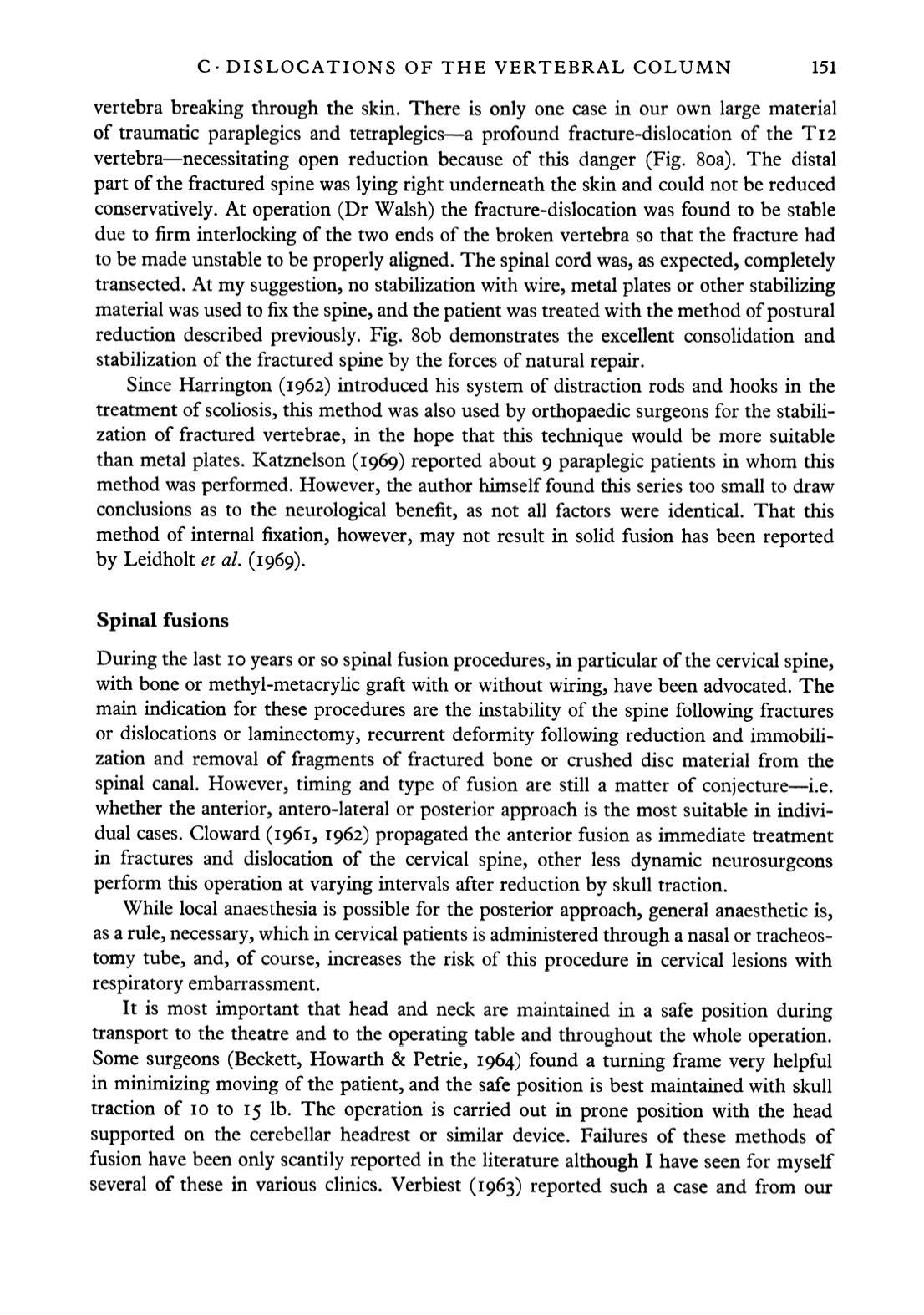
vertebra breaking through the skin. There is only one case in our own large material
of traumatic paraplegics and tetraplegics—a profound fracture-dislocation of the Ti2
vertebra—necessitating open reduction because of this danger (Fig. 8oa). The distal
part of the fractured spine was lying right underneath the skin and could not be reduced
conservatively. At operation (Dr Walsh) the fracture-dislocation was found to be stable
due to firm interlocking of the two ends of the broken vertebra so that the fracture had
to be made unstable to be properly aligned. The spinal cord was, as expected, completely
transected. At my suggestion, no stabilization with wire, metal plates or other stabilizing
material was used to fix the spine, and the patient was treated with the method of postural
reduction described previously. Fig. Sob demonstrates the excellent consolidation and
stabilization of the fractured spine by the forces of natural repair.
Since Harrington (1962) introduced his system of distraction rods and hooks in the
treatment of scoliosis, this method was also used by orthopaedic surgeons for the stabili
zation of fractured vertebrae, in the hope that this technique would be more suitable
than metal plates. Katznelson (1969) reported about 9 paraplegic patients in whom this
method was performed. However, the author himself found this series too small to draw
conclusions as to the neurological benefit, as not all factors were identical. That this
method of internal fixation, however, may not result in solid fusion has been reported
by Leidholt
el al.
(1969).
Spinal fusions
During the last 10 years or so spinal fusion procedures, in particular of the cervical spine,
with bone or methyl-metacrylic graft with or without wiring, have been advocated. The
main indication for these procedures are the instability of the spine following fractures
or dislocations or laminectomy, recurrent deformity following reduction and immobili
zation and removal of fragments of fractured bone or crushed disc material from the
spinal canal. However, timing and type of fusion are still a matter of conjecture—i.e.
whether the anterior, antero-lateral or posterior approach is the most suitable in indivi
dual cases. Cloward (1961, 1962) propagated the anterior fusion as immediate treatment
in fractures and dislocation of the cervical spine, other less dynamic neurosurgeons
perform this operation at varying intervals after reduction by skull traction.
While local anaesthesia is possible for the posterior approach, general anaesthetic is,
as a rule, necessary, which in cervical patients is administered through a nasal or tracheos-
tomy tube, and, of course, increases the risk of this procedure in cervical lesions with
respiratory embarrassment.
It is most important that head and neck are maintained in a safe position during
transport to the theatre and to the operating table and throughout the whole operation.
Some surgeons (Beckett, Howarth & Petrie, 1964) found a turning frame very helpful
in minimizing moving of the patient, and the safe position is best maintained with skull
traction of 10 to 15 Ib. The operation is carried out in prone position with the head
supported on the cerebellar headrest or similar device. Failures of these methods of
fusion have been only scantily reported in the literature although I have seen for myself
several of these in various clinics. Verbiest (1963) reported such a case and from our


