E • COMPLICATIONS
197
intermittent positive pressure ventilation as the commonest type (Johnston
et
a/., 1967;
Cooper & Grillo, 1969; and others). The stenosis at cuff level is of circumferential type,
leading to dense fibrous strictures. Tube-tip stenosis is the result of a long tracheostomy
tube impinging the lower portion of the trachea. This may result in ulceration of the
carina or affecting the right main bronchus leading to collapse of the right upper lobe or
collapse of the left lung. The backward inclination of the trachea and the curvature
of the tracheostomy tube may produce ulceration of the anterior wall and may lead to
perforation of the innonimate artery (Watts, 1963), while ulceration of the posterior
wall of the trachea may result in tracheo-oesophageal fistula.
It must be remembered that, in the acute stages of cervical lesions, as a result of the
paralysis of the vasoconstrictors the tone of the tissues in the trachea is lowered as it is in
other parts of the body, such as the urethra and bladder. Therefore, the vulnerability
of the tracheal mucosa is increased, especially if an inflatable polythene-type tracheostomy
tube is used which is not deflated frequently. This has the risk of damaging the mucosa
and developing a pressure sore, which soon becomes infected, resulting in tracheal
stenosis (Guttmann, 1970). Gibson (1967) considered over-inflation of the cuff, move
ment of the tracheostomy tube with each stroke of the respirator and secondary infection
as the main causes of stenosis, and Bannister and Braver, 1969, blamed the relatively
hard Trotex' tracheostomy tube for this complication. Frankel (1970) described in
detail 4 patients of tracheal stenosis in whom the plastic Tortex'—cuffed tube was used
in recent years, contrary to the soft rubber tubes which were used before at Stoke
Mandeville with satisfactory results. Two of the patients were C5 and C6 lesions, one
encephelomyelitis and one Li fracture with associated chest injury. The patient with
encephelomyelitis was operated 15 months after tracheostomy by Mr G.Grimshaw,
chest surgeon in Oxford, who dissected out the dense fibrotic stricture and approximated
the cut end of the trachea with interrupted sutures, but the patient died 5 days later due
to rupture of the innominate artery into the trachea. In another case (C6), the excision
of the stenosis one year after injury was successful. In the two remaining cases, dilatation
of the stenosis proved unsuccessful and a long term use of size 34 Chevalier-Jackson had
to be inserted, which in both cases was eventually successfully removed. Following these
experiences, the Tortex' tube was abandoned and only rubber cuffed tubes are used as a
routine. The conservative treatment consists of repeated dilatation of the stenosis.
However, this should be carried out with greatest care and gentleness, as there is the
great risk of eliciting undesirable autonomic hyperreflexia leading to cardiac arrest,
which will be discussed in detail in the following paragraph.
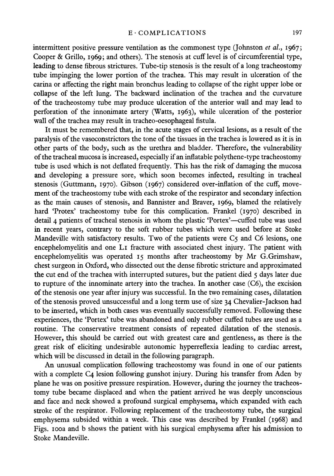
An unusual complication following tracheostomy was found in one of our patients
with a complete C4 lesion following gunshot injury. During his transfer from Aden by
plane he was on positive pressure respiration. However, during the journey the tracheos
tomy tube became displaced and when the patient arrived he was deeply unconscious
and face and neck showed a profound surgical emphysema, which expanded with each
stroke of the respirator. Following replacement of the tracheostomy tube, the surgical
emphysema subsided within a week. This case was described by Frankel (1968) and
Figs, icoa and b shows the patient with his surgical emphysema after his admission to
Stoke Mandeville.


