52
CHAPTER 7
gelatinous and consists of mucoid material, but by the end of the first decade a gradual
replacement of the mucoid material takes place by fibrocartilage originating from the
annulus fibrosis and from the covering cartilagenous plates of the upper and lower
surfaces of the vertebrae (Walmsley, 1950; Tondury, 1958). Its differentiation from the
annulus fibrosis is then less distinct and in due course it becomes amorphous and loses
its elasticity (Piischel, 1930). Although the anatomy of the intervertebral discs was
known for many centuries it is only during the last 40 years or so that their importance
in the pathology of the spine in relation to the pathology of the spinal cord and spinal
roots has been appreciated (Schmorl, 1927-32; Galland, 1930; Mixter & Barr, 1934;
Bradford & Spurling, 1945; Armstrong, 1957; and Frykholm, 1970).
The shape of the intervertebral discs is adapted to that of the vertebral bodies but
its thickness varies. While their thickness is fairly uniform in the thoracic region it is
increased in the cervical and lumbar regions, more anteriorly and posteriorly so that the
nucleus pulposus lies eccentrically and more close to the posterior longitudinal ligament.
Therefore, it is exposed to stresses such as distortion and tearing caused by fractures and
dislocations of vertebral bodies but also by occupational stress, lifting and carrying of
heavy weights, sportive activities, etc.
The discs are avascular apart from the supply of their peripheral parts from adjacent
blood vessels and they receive their nutrition from the spongy bone of the upper and
lower surface of the vertebrae.
LIGAMENTS
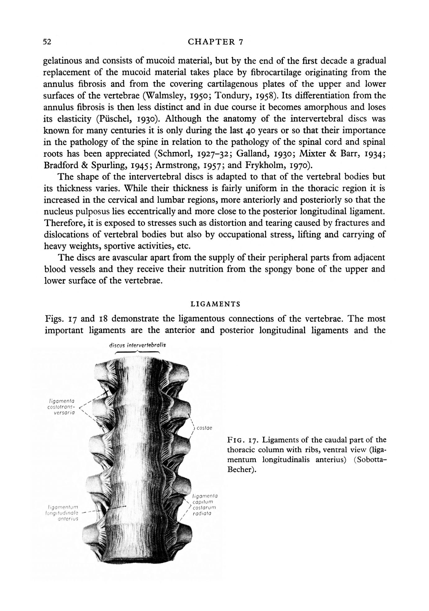
Figs. 17 and 18 demonstrate the ligamentous connections of the vertebrae. The most
important ligaments are the anterior and posterior longitudinal ligaments and the
//gomenfo
cos/o/rons-
versaria
ligamentum
longitudinal?
anterius
discus
/nferverfebro/j's
cosfoe
FIG. 17. Ligaments of the caudal part of the
thoracic column with ribs, ventral view (liga–
mentum longitudinalis anterius)
(Sobotta-
Becher).
/gomenfo
\ cop/'um
coslorum
rodiofo


