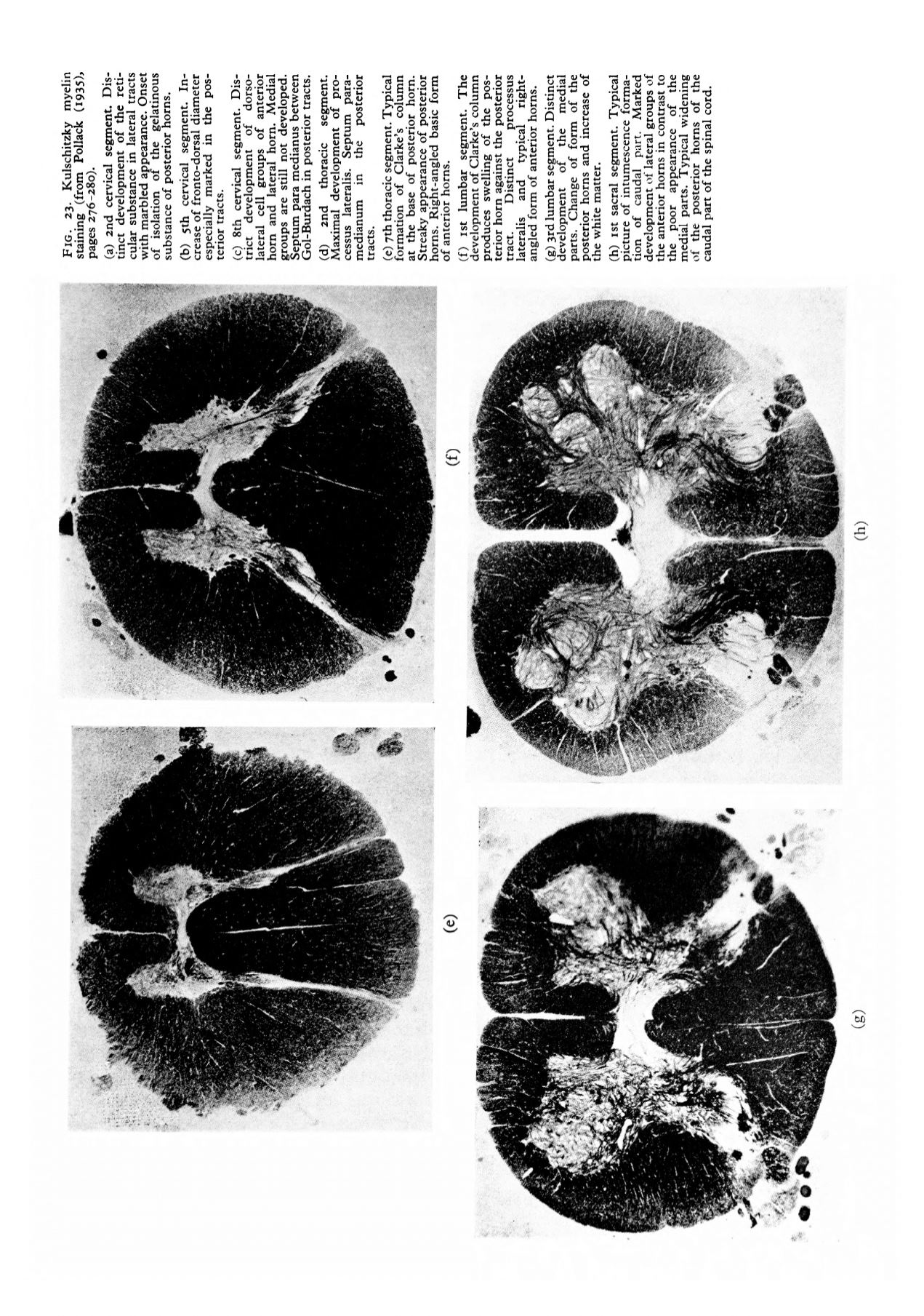
FIG. 23. Kulschitzky myelin
staining (from Pollack (i935)>
pages 276-280).
(a) 2nd cervical segment. Dis–
tinct development of the reti-
cular substance in lateral tracts
with marbled appearance. Onset
of isolation of the gelatinous
substance of posterior horns.
(b) 5th cervical segment. In–
crease of fronto-dorsal diameter
especially marked in the pos–
terior tracts.
(c) 8th cervical segment. Dis–
trict development of dorso-
lateral cell groups of anterior
horn and lateral horn. Medial
groups are still not developed.
Septum para medianus between
Gol-Burdach in posterior tracts.
(d)
2nd
thoracic
segment.
Maximal development of pro-
cessus lateralis. Septum para-
medianum
in
the
posterior
tracts.
(e) yth thoracic segment. Typical
formation of Clarke's column
at the base of posterior horn.
Streaky appearance of posterior
horns. Right-angled basic form
of anterior horns.
(f) ist lumbar segment. The
development of Clarke's column
produces swelling of the pos–
terior horn against the posterior
tract.
Distinct
processus
lateralis
and
typical
right-
angled form of anterior horns.
(g) 3rd lumbar segment. Distinct
development
of
the medial
parts. Change of form of the
posterior horns and increase of
the white matter.
(h) ist sacral segment. Typical
picture of intumescence forma–
tion of caudal part. Marked
development of lateral groups of
the anterior horns in contrast to
the poor appearance of
the
medial parts. Typical widening
of the posterior horns of the
caudal part of the spinal cord.


