66
CHAPTER 7
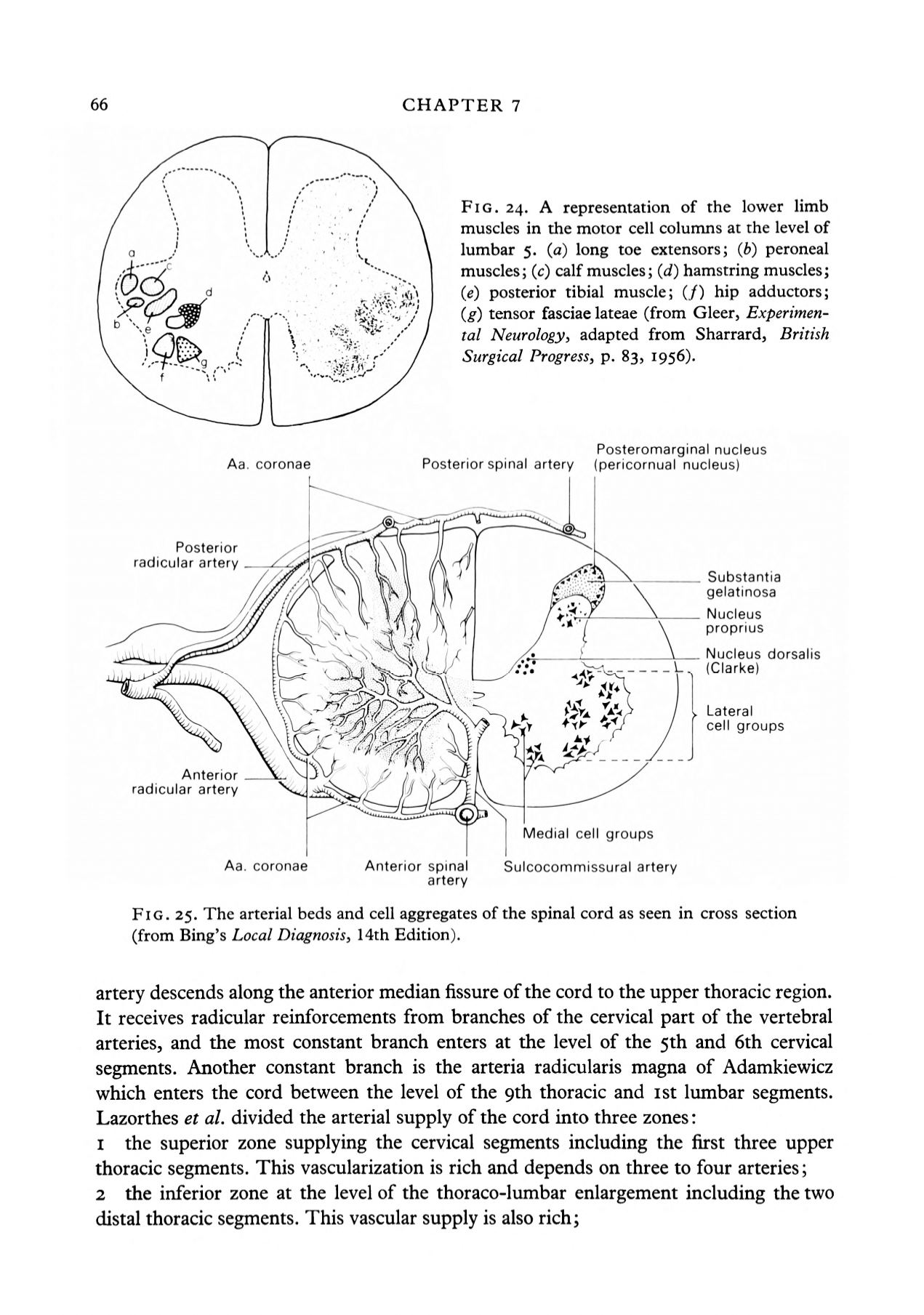
FIG. 24. A representation of the lower limb
muscles in the motor cell columns at the level of
lumbar 5.
(a)
long toe extensors;
(b}
peroneal
muscles;
(c)
calf muscles;
(d)
hamstring muscles;
(e)
posterior tibial muscle; (/) hip adductors;
(g)
tensor fasciae lateae (from Gleer,
Experimen
tal Neurology,
adapted from Sharrard,
British
Surgical Progress,
p. 83, 1956).
Aa. coronae
Posterior spinal artery
Posteromarginal nucleus
(pericornual nucleus)
Posterior
radicular artery
______\
Nucleus
-
___
\
(Clarke)
Substantia
gelatinosa
Nucleus
proprius
dorsalis
i r*i~.i.
~\
>
Lateral
cell groups
Anterior
radicular artery
Medial cell groups
coronae
Anterior spinal
Sulcocommissural artery
artery
FIG. 25. The arterial beds and cell aggregates of the spinal cord as seen in cross section
(from Bing's
Local Diagnosis,
14th Edition).
artery descends along the anterior median fissure of the cord to the upper thoracic region.
It receives radicular reinforcements from branches of the cervical part of the vertebral
arteries, and the most constant branch enters at the level of the 5th and 6th cervical
segments. Another constant branch is the arteria radicularis magna of Adamkiewicz
which enters the cord between the level of the 9th thoracic and ist lumbar segments.
Lazorthes
et al.
divided the arterial supply of the cord into three zones:
1
the superior zone supplying the cervical segments including the first three upper
thoracic segments. This vascularization is rich and depends on three to four arteries;
2
the inferior zone at the level of the thoraco-lumbar enlargement including the two
distal thoracic segments. This vascular supply is also rich;


