B ANATOMY, NEUROPATHOLOGY AND REGENERATION
75
artery as seen in C6 and 5 segments. The upwards spread of the cord damage is shown
in 4 segment by haemorrhages in the left lateral tract (Fig. 32). There is also a 'core' to be
seen in the upper ventral part of the left posterior column, which is also present though
small in C5 and C6 segments. The downwards spread of the damage is seen in
Cj
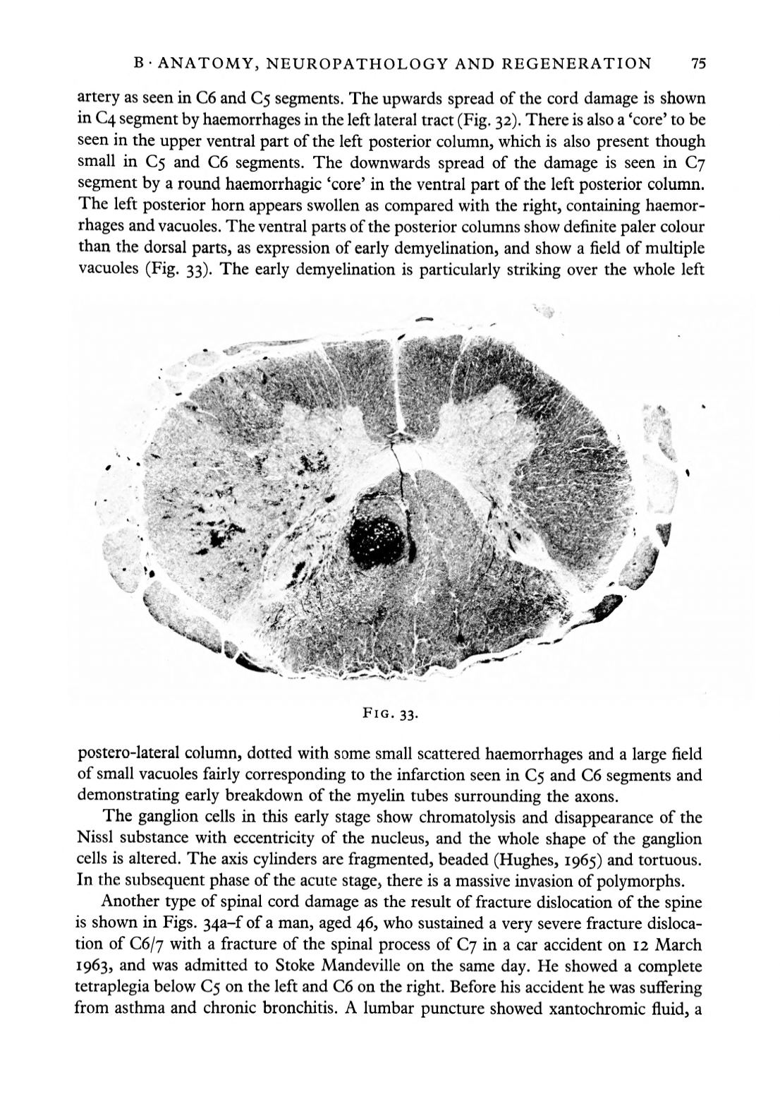
segment by a round haemorrhagic 'core' in the ventral part of the left posterior column.
The left posterior horn appears swollen as compared with the right, containing haemor–
rhages and vacuoles. The ventral parts of the posterior columns show definite paler colour
than the dorsal parts, as expression of early demyelination, and show a field of multiple
vacuoles (Fig. 33). The early demyelination is particularly striking over the whole left
FIG. 33.
postero-lateral column, dotted with some small scattered haemorrhages and a large field
of small vacuoles fairly corresponding to the infarction seen in C5 and C6 segments and
demonstrating early breakdown of the myelin tubes surrounding the axons.
The ganglion cells in this early stage show chromatolysis and disappearance of the
Nissl substance with eccentricity of the nucleus, and the whole shape of the ganglion
cells is altered. The axis cylinders are fragmented, beaded (Hughes, 1965) and tortuous.
In the subsequent phase of the acute stage, there is a massive invasion of polymorphs.
Another type of spinal cord damage as the result of fracture dislocation of the spine
is shown in Figs. 34a-f of a man, aged 46, who sustained a very severe fracture disloca–
tion of C6/7 with a fracture of the spinal process of
Cj
in a car accident on 12 March
1963, and was admitted to Stoke Mandeville on the same day. He showed a complete
tetraplegia below 5 on the left and C6 on the right. Before his accident he was suffering
from asthma and chronic bronchitis. A lumbar puncture showed xantochromic fluid, a


