68
CHAPTER 7
the cervical and lumbar regions, there was a marked preponderance on the left in the
thoracic region where the majority of the vessels entered, the sites of entry of the great
spinal arteries being between T8 and Tn on the left.
From the central anterior spinal artery sulcocommissural branches spring which pass
into the anterior median fissure and one passes alternatively to the right and left to
vascularize the anterior, lateral and also the anterior part of the posterior horn of the
grey matter but also part of the lateral and anterior white tracts (Mair & Druckman,
1953). The capillary network is much more dense in the grey than in the white matter
which explains its greater vulnerability following fractures and dislocations of the spine.
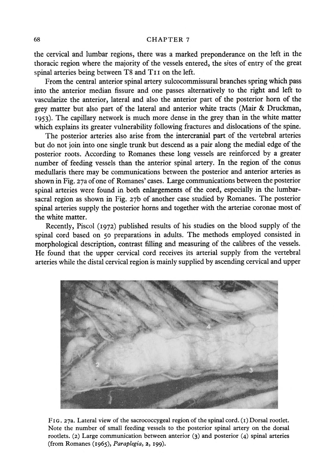
The posterior arteries also arise from the intercranial part of the vertebral arteries
but do not join into one single trunk but descend as a pair along the medial edge of the
posterior roots. According to Romanes these long vessels are reinforced by a greater
number of feeding vessels than the anterior spinal artery. In the region of the conus
medullaris there may be communications between the posterior and anterior arteries as
shown in Fig. 2ya ofone of Romanes' cases. Large communications between the posterior
spinal arteries were found in both enlargements of the cord, especially in the lumbar-
sacral region as shown in Fig.
2jb
of another case studied by Romanes. The posterior
spinal arteries supply the posterior horns and together with the arteriae coronae most of
the white matter.
Recently, Piscol (1972) published results of his studies on the blood supply of the
spinal cord based on 50 preparations in adults. The methods employed consisted in
morphological description, contrast filling and measuring of the calibres of the vessels.
He found that the upper cervical cord receives its arterial supply from the vertebral
arteries while the distal cervical region is mainly supplied by ascending cervical and upper
FIG. 273. Lateral view of the sacrococcygeal region of the spinal cord, (i) Dorsal rootlet.
Note the number of small feeding vessels to the posterior spinal artery on the dorsal
rootlets. (2) Large communication between anterior (3) and posterior (4) spinal arteries
(from Romanes (1965),
Paraplegia,
2, 199).


