B • ANATOMY, NEUROPATHOLOGY AND REGENERATION
73
the pia-arachnoidea haemorrhagic and there are also patches of haemorrhages on the
inside of the dura. The line of the long spinal artery is interrupted.
Microscopically, the first stage is characterized by haemorrhages involving very
extensively the highly vascularized and spongious and therefore most vulnerable grey
substance, especially in the centre but also spreading to varying degrees into the white
matter.
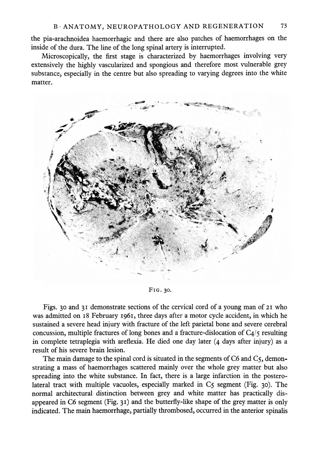
FIG. 30.
Figs. 30 and 31 demonstrate sections of the cervical cord of a young man of 21 who
was admitted on 18 February 1961, three days after a motor cycle accident, in which he
sustained a severe head injury with fracture of the left parietal bone and severe cerebral
concussion, multiple fractures of long bones and a fracture-dislocation of 04/5 resulting
in complete tetraplegia with areflexia. He died one day later (4 days after injury) as a
result of his severe brain lesion.
The main damage to the spinal cord is situated in the segments of C6 and C5, demon–
strating a mass of haemorrhages scattered mainly over the whole grey matter but also
spreading into the white substance. In fact, there is a large infarction in the postero-
lateral tract with multiple vacuoles, especially marked in 5 segment (Fig. 30). The
normal architectural distinction between grey and white matter has practically dis–
appeared in C6 segment (Fig. 31) and the butterfly-like shape of the grey matter is only
indicated. The main haemorrhage, partially thrombosed, occurred in the anterior spinalis


