72
CHAPTER 8
sense. It is probable that in these cases some petechial haemorrhages, followed by oedema,
may have been the cause of the delay in functional recovery. It must be admitted that
the underlying mechanism of spinal cord concussion is still obscure. However, in the
light of the modern advances made on the interneurone function of synapses and their
afferent and efferent connections and the chemical transmission between neurones, one
cannot help feeling that Obersteiner's concept of the 'molecular disturbance of neurones'
may be after all an acceptable explanation of the mechanism involved in spinal cord
concussion.
CONTUSION, LACERATION, TRANSECTION
Contusion, the most frequent type of injury of the spinal cord, produces a great variety
of detectable organic alterations in the axons, parenchyma, vascular supply and meninges
which may be combined or independent of each other. The damage to the spinal cord is
caused by the impact of the violence, leading to acute compression of the cord and
resulting in destruction of the nervous elements and their blood supply at the site of the
main impact of the violence but also producing pressure, squeezing and stretch effects
on several segments above and below the main injury. The alterations in the tissues con–
sist of petechial haemorrhages due to rhexis or diapedesim, haematomyelia, thrombosis,
resulting in softening of the parenchyma with demyelination and necrosis, cyst formation
and fibrotic and glial scarring. At some stage, there is proliferation of vessels and glial
cell proliferation. Several authors (Kienbeck, 1902; Klaue, 1969; Mayer & Peters, 1971)
have drawn special attention to vacuolization in the white matter of the cord, forming
distinct fields of vacuoles (Liickenfelder). They represent the breaking down of the
myelin tubes surrounding the axons, which themselves undergo chromatolysis, and the
disappearance of the Nissl bodies from the centre of the cell.
Three stages in the pathological changes of the cord as a result of contusion can be
distinguished, although the transition from one to another is rather fluent.
Acute stage
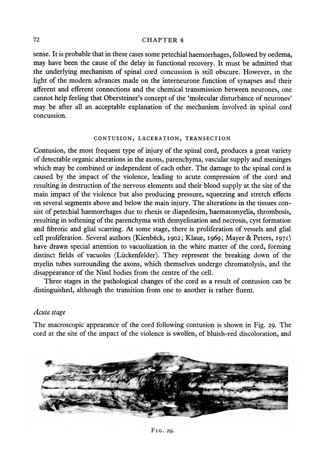
The macroscopic appearance of the cord following contusion is shown in Fig. 29. The
cord at the site of the impact of the violence is swollen, of bluish-red discoloration, and
FIG. 29.


