E • COMPLICATIONS
211
of the paralysed leg or forearm is diminished. X-ray investigation at this stage reveals
cloudy, streaky or patchy densities in the muscles involved.
2
Stage of calcification. This can develop within a few weeks and even within a few
days (Rosak, 1961). The X-ray shows irregularly shaped calcareous deposits within the
para-articular tissues.
3
Stage of ossification. The process of ossification is finalized and has led to dense
ossification of ligaments, fasciae and muscles surrounding the joints, resulting in extra-
articular ankylosis. At operation, the bony mass is often found to be quite eburnean.
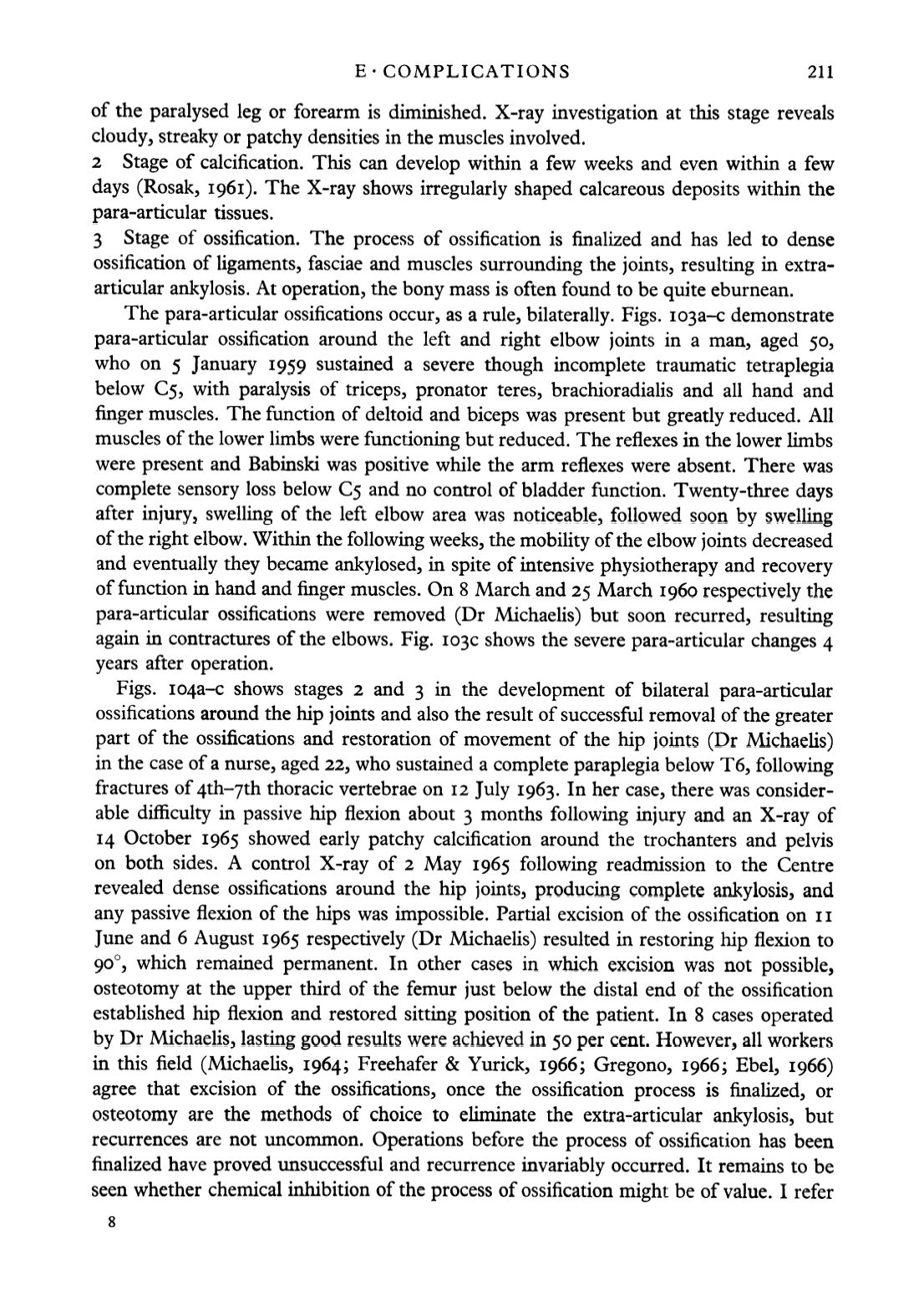
The para-articular ossifications occur, as a rule, bilaterally. Figs. i03a-c demonstrate
para-articular ossification around the left and right elbow joints in a man, aged 50,
who on 5 January 1959 sustained a severe though incomplete traumatic tetraplegia
below 05, with paralysis of triceps, pronator teres, brachioradialis and all hand and
finger muscles. The function of deltoid and biceps was present but greatly reduced. All
muscles of the lower limbs were functioning but reduced. The reflexes in the lower limbs
were present and Babinski was positive while the arm reflexes were absent. There was
complete sensory loss below €5 and no control of bladder function. Twenty-three days
after injury, swelling of the left elbow area was noticeable, followed soon by swelling
of the right elbow. Within the following weeks, the mobility of the elbow joints decreased
and eventually they became ankylosed, in spite of intensive physiotherapy and recovery
of function in hand and finger muscles. On 8 March and 25 March 1960 respectively the
para-articular ossifications were removed (Dr Michaelis) but soon recurred, resulting
again in contractures of the elbows. Fig. I03C shows the severe para-articular changes 4
years after operation.
Figs. I04a-c shows stages 2 and 3 in the development of bilateral para-articular
ossifications around the hip joints and also the result of successful removal of the greater
part of the ossifications and restoration of movement of the hip joints (Dr Michaelis)
in the case of a nurse, aged 22, who sustained a complete paraplegia below T6, following
fractures of 4th-yth thoracic vertebrae on 12 July 1963. In her case, there was consider
able difficulty in passive hip flexion about 3 months following injury and an X-ray of
14 October 1965 showed early patchy calcification around the trochanters and pelvis
on both sides. A control X-ray of 2 May 1965 following readmission to the Centre
revealed dense ossifications around the hip joints, producing complete ankylosis, and
any passive flexion of the hips was impossible. Partial excision of the ossification on 11
June and 6 August 1965 respectively (Dr Michaelis) resulted in restoring hip flexion to
90°, which remained permanent. In other cases in which excision was not possible,
osteotomy at the upper third of the femur just below the distal end of the ossification
established hip flexion and restored sitting position of the patient. In 8 cases operated
by Dr Michaelis, lasting good results were achieved in 50 per cent. However, all workers
in this field (Michaelis, 1964; Freehafer & Yurick, 1966; Gregono, 1966; Ebel, 1966)
agree that excision of the ossifications, once the ossification process is finalized, or
osteotomy are the methods of choice to eliminate the extra-articular ankylosis, but
recurrences are not uncommon. Operations before the process of ossification has been
finalized have proved unsuccessful and recurrence invariably occurred. It remains to be
seen whether chemical inhibition of the process of ossification might be of value. I refer


