F- CLINICAL ASPECTS OF SPINAL CORD INJURIES
395
large diverticulum resulting from the stone at the peno-scrotal junction. Fig. 171 shows
a collection of the various types and sizes of stones encountered throughout the years at
Stoke Mandeville. Special attention is drawn to the two large bladder stones, one lying
on top of the other like millstones, found in a paraplegic woman and successfully removed
by Dr Walsh. It is interesting to note that these overriding stones caused grinding
noises in the patient's lower abdominal region, but when the patient reported hearing
these noises, she was promptly treated with tranquillizers! (This case who later became
pregnant has been described on page 192.)
Stones in paraplegic patients consist of phosphates and carbonates of calcium,
magnesium and ammonium. Their chemical properties have been studied in detail, in
particular by Mulvaney (1964) and recently by Burr (1972), the biochemist of Stoke
Mandeville Spinal Centre. He employed for the analysis of stones the electron microprobe
method used by geologists to examine rock samples, and Figs. 172, 173 show a section
of a ureteric stone and the tracings obtained by this section for calcium, magnesium and
phosphorus. Of particular interest is the part marked with an arrow in the tracings at
which the calcium concentration falls almost to zero but the magnesium concentration
is so high that the tracing is off the scale, while there is no apparent change in the phos
phorus concentration. Although the original stone nucleus in a few paraplegic patients
may be urate, oxalate and even cystine, the secondary stone consists of calcium phosphate
in the great majority of cases. Amongst our paraplegics, there was one patient who, before
his paraplegia, had an oxalate stone, and when he developed a larger stone following his
spinal injury the stone nucleus was oxalate.
The incidence of calculosis following spinal cord injuries has been repeatedly reported
in the literature, and from the statistics published in this country, Canada and U.S.A.
during and in the early years after the Second World War, which were compiled in my
monograph of 1953, tne incidence of calculosis varied between 20-68-7 Per cent - My
own statistic of 351 traumatic paraplegics from the Second World War revealed a calcu
losis of 22-5 per cent. In the great majority of these patients, the initial treatment had
been suprapubic cystostomy. The considerable improvement made ever since in the
management of paraplegics and tetraplegics resulting in prevention of calculosis, is best
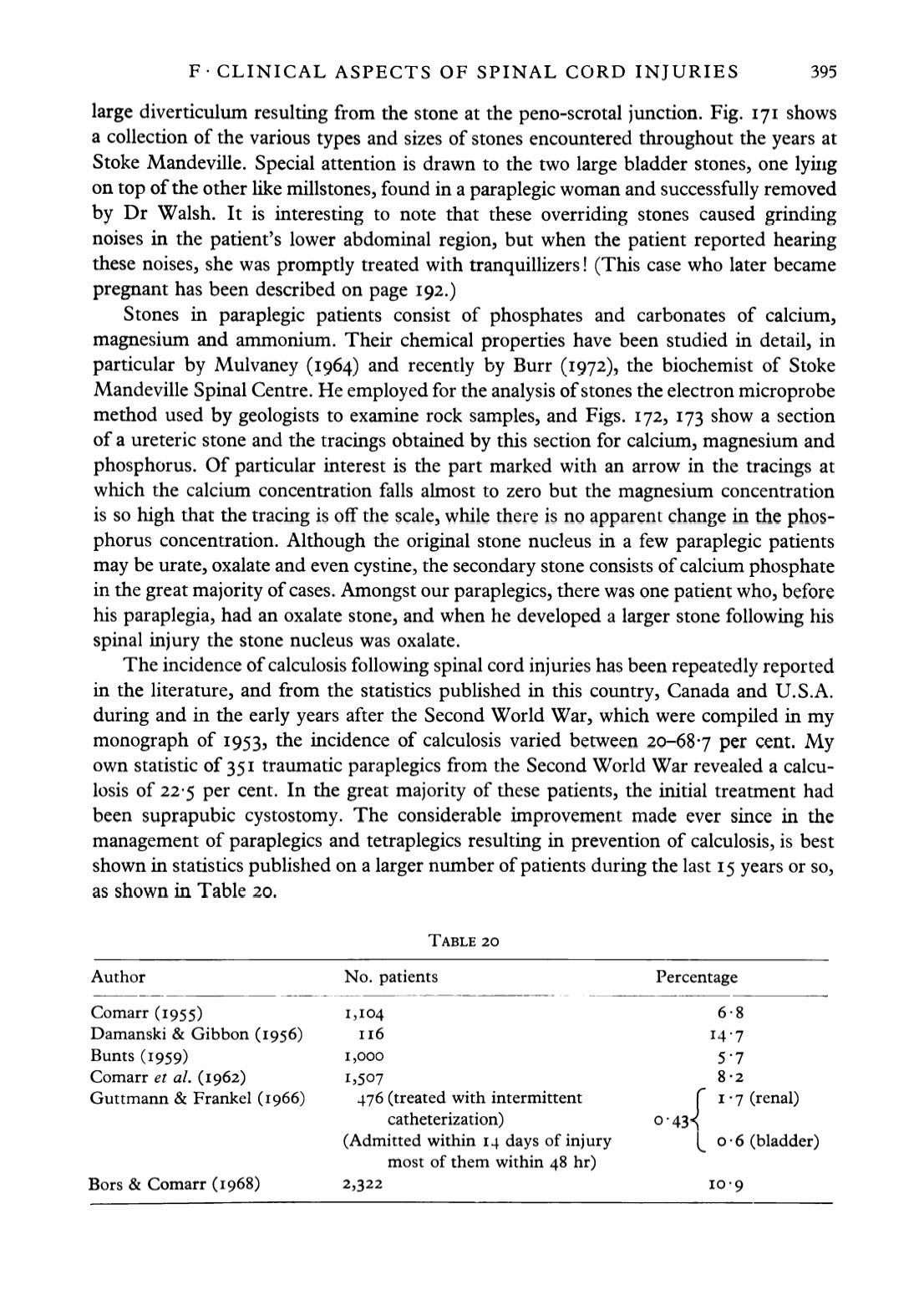
shown in statistics published on a larger number of patients during the last 15 years or so,
as shown in Table 20.
TABLE 20
Author
No. patients
Percentage
Comarr (1955)
1^104
6-8
Damanski & Gibbon (1956)
116
14-7
Bunts (1959)
1,000
5-7
Comarr
et al.
(1962)
1,507
8-2
Guttmann & Frankel (1966)
476 (treated with intermittent
C
1-7 (renal)
catheterization)
0-43^
(Admitted within 14 days of injury
^ 0-6 (bladder)
most of them within 48 hr)
Bors & Comarr (1968)
2,322
10-9


